Wilson’s
disease (hepatolenticular degenera-tion) is an autosomal recessive disease of
copper metabolism due to mutation in ATP7B gene1,2. The genetic
defect causes excessive copper accumulation in the liver, brain and other body
tissues.
The
prevalence of Wilson’s disease is one in 30,000 people worldwide and
corresponding carrier frequency is one in 903. Clinically, it
presents as liver disease or neurological / neuropsychiatric disorder in
different age groups (Table 1)4. It manifests as liver disease in
children and young adults, typically between the ages of 6 and 45 years.
Neurological and psychiatric symptoms are seen in adults in their twenties and
older5,6.
The
identification of Kayser – Fleischer ring is helpful in the diagnosis of
Wilson’s disease. Patients suspected of this disease are referred to ophthalmo-logist
for identification of Kayser – Fleischer ring by slit-lamp examination and
gonioscopy. It is a rare disease and few ophthalmologists have ever seen a true
Kayser – Fleischer ring. It is reported that often the Kayser – Fleischer rings
of one patient are seen by multiple ophthalmologists in the department, so the
total number of patients diagnosed is less than the total number of reported
cases seen7.
We present a case of Wilson’s
disease with neurological manifestations and Kayser – Fleischer ring without
chronic hepatic involvement. This case has classic clinical features of the
disease and typical Kayser – Fleischer rings in the cornea. The ophthalmo-logist
has an important role in identification of this disease in suspected cases but
he is not directly involved in the treatment of such cases. It is considered
appropriate to report this case and discuss clinical features, current status
of management and prognosis of the disease so that they feel confident in
management of this disease.
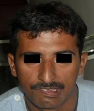
Fig. 1: A mask like facies with a vacuous smile, dysarthria and
bradykinesia.

Fig. 2: Arrows indicate greenish - brown Kayser Fleisher Ring in
descemet membrane of cornea.
CASE REPORT
A male
patient, 31 years of age, presented with two weeks history of difficulty in
speaking and tremors of hands. He was married and had two healthy daughters.
His parents were alive and healthy. He was conscious, well oriented and had
stable vital signs. Neurological examination revealed mask like facies with a
vacuous smile, dysarthria and bradykinesia (Fig 1). Kayser - Fleisher rings
were seen on slit lamp examination (Fig. 2). There was no clinical evidence of
chronic liver disease.
The laboratory investigations
showed haemo-globin 13.9
g/dl, platelet count 161 × 109/l, WBC 6.5 × 10⁹/l, serum
ALT 17 U/l (9 - 43 U/l), serum alkaline phosphatase 332 U/l (80 - 306 U/l),
total bilirubin 23 umol/l (< 19 umol/l), urea 4.4 mmol/l (3.2 - 6.7 mmol/l)
and creatinine 105 umol/l (53 - 120 umol/l). Serum ceruloplasmin was 10 mg/dl
(19 - 57 mg/dl). MRI brain showed hyper intense signals in caudate nuclei,
lentiform nuclei, thalami and brainstem on T2W images and FLAIR (Fig. 3). A
diagnosis of Wilson’s disease was made and Penicillamine (Vistamine) with oral
Zinc was started. Follow up after 3 months showed improvement in clinical
features, serum alkaline phosphatase 309 U/L (80 - 306 U/l), total bilirubin 21
(< 19 umol/l), urea 4.3 mmol/l (3.2 - 6.7 mmol/l) and creatinine 96 umol/l
(53 - 120 umol/l) and serum ceruloplasmin was 11.9 mg/dl
(19 - 57 mg/dl). Follow up planned at 6 months and 12 months after treatment.
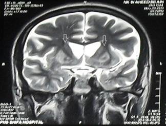
Fig. 3: MRI brain shows
hyperintense signals in caudate nuclei, lentiform nuclei, thalami and brainstem
on T2 W Images and FLAIR.
DISCUSSION
Samuel
Alexander Kinnier Wilson (1878 - 1937) described this condition in 1912. The
neurological form of Wilson’s disease is also known as Westphal - Strumpell
pseudosclerosis.
Table 1:
Clinical
features in Wilson’s disease 3
|
Hepatic ·
Asymptomatic hepatomegaly ·
Splenomegaly ·
Persistently elevated serum
aminotransferase activity (AST, ALT) ·
Fatty liver ·
Acute hepatitis / Resembling autoimmune
hepatitis ·
Cirrhosis: compensated or decompensated ·
Acute liver failure |
|
Neurological ·
Movement disorders (tremor, involuntary
movements) ·
Drooling, Dysarthria ·
Rigid dystonia ·
Pseudobulbar palsy ·
Dysautonomia ·
Migraine headaches ·
Seizures |
|
Psychiatric ·
Depression ·
Neurotic behaviours / Psychosis ·
Personality change |
|
Other symptoms ·
Ocular: Kayser Fleischer rings, sunflower
cataracts ·
Cutaneous: lunulae ceruleae ·
Renal abnormalities: aminoaciduria &
nephrolithiasis ·
Skeletal abnormalities: premature
osteoporosis and arthritis ·
Pancreatitis ·
Hypothyroidism ·
Menstrual irregularities: infertility,
repeated miscarriages |
Wilson’s disease manifests as
neurological disease in adults8. It can present as movement
disorders or rigid dystonias. Movement disorders appear earlier as tremors,
poor coordination, loss of fine-motor control, micrographia, chorea and / or
choreoathetosis. Spastic dystonia disorder manifests as mask-like facies,
rigidity and gait disturbances5. Pseudobulbar involve-ment is more
common in older individuals and presents as dysarthria, drooling and difficulty
in swallowing.
Table 2:
Diagnostic tests of Wilson’s Disease 12
|
·
Kayser-Fleischer Rings ·
Low Serum Ceruloplasmin levels (<0.20
g/l, normal is 0.20 to 0.40 g/l) ·
24 hour Urinary Copper Excretion (>100
µg/day or 1.0 mol/day) ·
24 hour Urinary Copper Excretion after
D-penacillamine (>25 mol/day) ·
Hepatic Copper Level on liver biopsy
(>250 µg/g dry weight, normal is< 50 µg/g dry weight) ·
Genetic mutation in ATP7B gene |
The
neurologic findings in patients with hepatic presentation may be subtle. Mood
disturbance, depression and changes in school performance may be observed5.
The psychiatric manifestations are variable. Pure psychotic disorders are
uncommon.
The
Kayser – Fleischer ring is the hall mark of Wilson’s disease9. The
copper deposition in Descemet’s membrane of the cornea appears as
Kayser-Fleischer ring and indicates a high level of copper in the body10.
It appears as a golden brown ring in the peripheral cornea, extending from
Schwalbe’s line to less than 5 mm on to the cornea. It can be greenish yellow,
ruby red or bright green in colour. It is almost always bilateral. Initially,
it appears superiorly, then inferiorly, and later becomes circumferential.
Gonioscopy is often required in early stages of disease but it can be seen in
torch light in advanced disease. It is seen in about 85 – 100% patients with
neurological and/or psychiatric manifestations, 33 – 86% patients with hepatic
disease and up to 59% in asymptomatic patients11. With treatment, it
disappears in 85 – 90% of patients12,13. Sunflower cataract appears
as a late manifestation of neurological form of Wilson’s disease. In torch
light, it appears as greenish disc in the centre of the pupil and on slit – lamp
examination, it appears as brown / green pigmentation of the anterior and
posterior lens capsule14.
The
presence of Kayser-Fleischer rings, neurolo-gical symptoms and low serum
ceruloplasmin are considered diagnostic of Wilson’s disease15.
Further tests are advised where indicated (Table 2)7. Wilson’s
disease is suspected in close relatives of the patient and relevant clinical
feature. In neurolocial symptoms, MRI brain shows hyperintensities in the basal
ganglia in the T2 setting. It may show the characteristic 'Face of the
giant panda’ sign16.
Liver
biopsy is the gold standard test and more than 250ug of copper per gram of
dried liver tissue confirms Wilson’s disease. Mutation analysis of the ATP7B
gene may be performed1,2. If confirmed, family members can be
screened as part of clinical genetic family counseling.
These
patients are advised to take a diet low in copper – containing foods and avoid
mushrooms, nuts, chocolates, dried fruits, liver and shell fish. They need a
life-long treatment and it should not be discontinued. Symptomatic patients are
treated with chelating agents17,18. Penicillamine is advised as
tablet D-penicillamine by mouth 2 or 3 times a day19. Pyridoxine
must be given with it. Full blood count and urinalysis is monitored regularly.
24 hours urinary copper values should be 5 – 10 times normal to confirm
chelation and increased urinary excretion of copper. Lower values suggest
non-compliance or body stores may have been adequately depleted. Serious side
effects are seen in up to 30% patients and include severe thrombocytopenia,
leucopenia, aplastic anemia, proteinuria, nephritic syndrome, polyserositis,
Goodpasture syndrome and severe skin reactions. If side effects occur, tablet
D-penicillamine is substituted with tablet trientine hydrochloride as an alternate
treatment. If it is not available, these adverse events might be manageable
with co-administration of steroids. Almost 50% patients with neurological
disorder experience a paradoxical worsening in their symptoms with
penicillamine.
Once
laboratory investigations are within normal limits, zinc therapy is given to
maintain stable copper levels20. Tablet Zinc acetate (Galzin) is
advised at least 2 – 3 time daily before meals. It stimulates metallo-thionein
which is a protein in gut cells that binds copper and prevents its absorption
and transport to the liver.
The Kayser – Fleischer rings
can be identified accurately by an ophthalmologist by using a slit lamp. The
presence of Kayser-Fleischer rings, neurological symptoms and low serum
ceruloplasmin are helpful in diagnosis of Wilson’s disease. A high index of
suspicion is required for early detection of Wilson’s disease in adolescents
and young adults with neurological disorders. Initiation of treatment at an
early stage can prevent complications.
Author’s Affiliation
Dr. Hannan Masud
Classified Ophthalmologist
CMH Pano Aqil
Sindh
Dr. Tariq Bashir
Classified Medical Specialist
CMH Pano Aqil
Sindh
REFERENCES
1.
Gromadzka
G, Schmidt HH, Genschel J, Bochow B, Rodo M, Tamacka B, Litwin T, Chabik G,
Czlonkowska A. p.H1069Q mutation in ATP7B and biochemical parameters of copper
metabolism and clinical manifestation of Wilson’s disease. Mov Disord. 2006;
21: 245-8.
2.
Huster D,
Kühne A,
Bhattacharjee A,
Raines L,
Jantsch V,
Noe J, Schirrmeister W,
Sommerer I,
Sabri O,
Berr F,
Mössner J,
Stieger B,
Caca K,
Lutsenko S. Diverse functional properties of Wilson disease ATP7B
variants. Gastroenterology. 2012; 142: 947-56.
3.
Olivarez
L, Caggana M, Pass KA, Ferguson P, Bremer GJ. Estimate
of the frequency of Wilson’s disease in the US Caucasian population: a mutation
analysis approach. Ann Hum Genet. 2001; 65: 459-63.
4.
Roberts
EA, Schilsky ML. Diagnosis and treatment of Wilson disease: An update. Hepatology.
2008; 47: 2089–211.
5.
Coffey
AJ, Durkie M, Hague S, McLay K, Emmerson J, Lo C, Klaffke S, Joyce CJ, Dhawan
A, Hadzic N, Mieli-Vergani G, Kirk R, Elizabeth Allen K, Nicholl D, Wong S,
Griffiths W, Smithson S, Giffin N, Taha A, Connolly S, Gillett GT, Tanner S,
Bonham J, Sharrack B, Palotie A, Rattray M, Dalton A, Bandmann O. A genetic study of Wilson's disease
in the United Kingdom. Brain. 2013; 136: 1476-87.
6.
Kumar
MK, Kumar V, Singh PK. Wilson’s Disease with Neurological Presentation, without Hepatic
Involvement in Two Siblings.
J Clin Diagn Res. 2013; 7: 1476-8.
7.
Birkholz
ES, OettingTA. Kayser-Fleischer Ring: A Systems Based Review of the
Ophthalmologist’s Role in the Diagnosis of Wilson’s Disease. Eye Rounds.otg.
2009.
8.
Svetel
M, Kozic D, Stefanova E, Semnic R, Dragasevic N, Kostic VS.
Dystonia in Wilson’s disease. Mov disord. 2001; 16: 719-23.
9.
Liu M,
Cohen EJ, Brewer GJ, Laibon PR. Kayser Fleischer ring as the
presenting sign of Wilson disease. Am J Ophthalmol. 2002; 133: 832-4.
10. Suvarna JC. Kayser-Fleischer
ring. J Postgrad Med. 2008; 54: 238-40.
11.
Mak CM,
Lam CW. Diagnosis of Wilson’s disease: a comprehensive review. Critical
Reviews in Clinical Laboratory Sciences. 2008; 45: 263-90.
12.
Lossner
A, Lossner J, Bachmann H, Zotter J. The Kayser Fleischer ring
during long-term treatment in Wilson’s disease (hepatolenticular degeneration):
A follow-up Study. Graefes Arch Clin Exp Ophthalmol. 1986; 224: 152-5.
13.
Fenu M,
Liggi M,
Demelia E,
Sorbello O,
Civolani A,
Demelia L. Kayser-Fleischer
ring in Wilson's disease: a cohort study. Eur J Intern Med. 2012; 23:
150-6.
14.
Cairns JE,
Williams HP,
Walshe JM.
Sunflower cataract in Wilson’s disease. Br Med J. 1969; 3: 95-6.
15.
Martins
da Costa C, Baldwin D, Portmann B, Lolin Y, Mowat AP, Mieli-Vergani G. Value
of urinary copper excretion after penicillamine challenge in the diagnosis of
Wilson’s disease. Hepatology. 1992; 15: 609-61.
16.
Kuruvilla
A, Joseph S. 'Face of the giant panda' sign in Wilson's disease revisited.
Neurol India. 2000; 48: 395.
17.
Roberts
EA, Schilsky ML. Diagnosis and treatment of Wilson’s disease: an update.
Hepatology. 2008; 47: 2089-111.
18.
European Association for Study of Liver; EASL Clinical Practice
Guidelines: Wilson’s disease. J Hepatol. 2012; 56: 671-85.
19.
Durand
F, Bernuau J, Giostra E, Mentha G, Shouval D, Degott C, Benhamou JP, Valla D.
Wilson’s disease with severe hepatic insufficiency: beneficial effects of early
administration of D-penicillamine. Gut. 2001; 48: 849-52.
20. Brewer GJ. Zinc
acetate for the treatment of Wilson’s disease. Expert Opin Pharmacother. 2001;
2: 1473-7.